Gastroenterology Clinic

Dr Arti Rattan has extensive clinical experience in diagnosing and treating problems with the digestive organs and liver disorders. The digestive organs include the digestive tract-oesophagus, stomach, small and large intestine-as well as related organs-the liver, pancreas, and gallbladder.

Inflammatory Bowel Disease (IBD)

Inflammatory bowel disease (IBD) is inflamation of the intestines. The cause is unknown however it’s believed to be a combination of genetic, environmental and  immunological factors. Triggers such as viruses, bacteria and/or proteins prompt the immune system to to switch on its normal defence mechanism (inflammation) against a foreign substance. With most people, this response goes away once the foreign substance is destroyed. In some people, the body’s reaction continues. Prolonged inflamation eventually damages the walls of the gastrointestinal tract.

Symptoms of IBD

Abdominal pain, vomiting, diarrhea, severe urgency, rectal bleeding, severe internal cramps/muscle spasms in the region of the pelvis, fatigue, weight loss and anaemia due to blood loss. For most people the symptoms come and go between periods of remission and flare-ups.

Treatment of IBD

IBD cannot be cured but it can be managed with medication, nutrition and dietetics therapies and antibiotics. Controlling the inflamation allows the tissue to heal and symptoms to ease.

Irritable Bowel Syndrome (IBS)

Irritable bowel syndrome is an intestinal disorder causing pain in the stomach, wind, diarrhoea and constipation. The cause is unknown however changes of routine, emotional stress, infection and food intolerance, can trigger an attack.

Symptoms of IBS

Abdominal pain, cramping, bloating (these can be relieved by passing wind or going to the toilet), alternating diarrhoea and constipation, mucus in stools, nausea.

Treatment of IBS

IBS cannot be cured but it can be managed with changes in lifestyle and diet, reviewing any medications, anti-diarrhoeal medicines, constipation treatments and antispasmodics (to ease cramping). Finding a therapist with experience in the successful treatment of IBS is important.

Bowel cancer screening

Bowel cancer is Australia’s second biggest cancer killer. It can be treated in up to 90 per cent of cases, early detection is the key. Bowel cancer begins when cells in the bowel lining grow too quickly. These growths are often benign, but over time, some growths become cancerous. If left untreated, cancers can develop and then spread to other parts of the body.

Both men and women are at risk of developing bowel cancer, however the risk increases if you:

  • are aged 50 years and over (your risk increases with age)
  • have had an inflammatory bowel disease (such as Crohn disease or ulcerative colitis)
  • have previously had special types of polyps, called adenomas, in the bowel
  • have a significant family history of bowel cancer or polyps

All Australians aged 50 and over are encouraged to have a bowel screening test done. The National Bowel Cancer Screening Program helps achieve this and to further prevent mortality secondary to bowel cancer. If your tests come back positive, it does not necessarily mean a diagnosis of colon cancer. It means that something that may be bleeding  needs to be investigated further. Most of the time causes are benign and easily treated e.g haemorrhoids or polyps. A GP specialist follow up is generally recommended after a positive FOBT.

Colonic Polyps

Colon polyps most frequently are benign (noncancerous) overgrowths on the surface of the colon. Most polyps are small, usually less than 1cm in size, although they can grow larger. Most polyps are harmless, but over time some can develop into bowel cancer. Because most bowel cancer begins as polyps, all polyps should be removed when found.

When signs and symptoms of colon polyps are present, they include:

  • Rectal bleeding
  • Anemia
  • Diarrhea
  • Constipation

Colonic Polyps treatment

Bowel polyps can be removed during a colonoscopy. Surgery is rarely needed to remove polyps, but may be necessary if the polyps are very large or can’t be reached safely during the colonoscopy. After removal, the polyp is sent to a pathology lab to check for any signs of cancer. The lab will also be able to tell if the polyp has been entirely removed or if there is a chance of it growing back.

If any signs of cancer are found, further treatment may be required.

Barrett’s Oesophagus

Gastroesophageal reflux disease (GERD) is a condition in which the stomach contents, including acid, reflux into the oesophagus. About 10 percent of patients with chronic GERD or inflammation of the oesophagus will develop Barrett’s oesophagus. Barrett’s oesophagus does not cause symptoms, although patients with Barrett’s oesophagus may have symptoms related to GERD. It does, though, increase the risk of developing oesophageal adenocarcinoma, which is a serious, potentially fatal cancer of the oesophagus.

Many people have no signs or symptoms, however if you have been experiencing the following for 2 weeks or more, it’s time to see a Gastroenterologist

  • Frequent heartburn or indigestion
  • Difficulty swallowing food
  • Blood in vomit or stool
  • Acidic or bitter liquid coming up to the chest or mouth during the night

If you’ve been diagnosed with Barrett’s esophagus, it’s important to have routine examinations. With these routine examinations, your doctor can discover precancerous and cancer cells early, before they spread and when the disease is easier to treat.

Barrett’s Oesophagus Procedures

Dr Arti Rattan will most likely perform an upper endoscopy with biopsy to help diagnose Barrett’s oesophagus. During this procedure, the lining of the oesophagus is checked for abnormalities. Endoscopy refers to the use of a flexible, lighted tube called an endoscope with a camera at the tip. The endoscope is inserted into your stomach through your mouth. There is a camera at the tip of the endoscope so your doctor can see any abnormal tissue. A small tissue sample is removed and sent to a pathology lab for analysis.

Barrett’s Oesophagus Treatment

If the diagnosis is confirmed the treatment may involve simple medication and lifestyle changes to help reduce acid and allow the damaged tissue to heal. Dr Arti Rattan will continue to monitor your condition to minimise the risk of the disease spreading or becoming cancerous. If there are precancerous cells (dysplasia) diagnosed in the Barrett’s oesophagus, Endoscopic Mucosal Resection treatment is recommended and proven safe and effective for preventing progression to cancer.

H pylori

H. pylori is a common type of bacteria that grows in the digestive tract and has a tendency to attack the stomach lining. It infects the stomachs of over half of the world’s adult population. H. pylori infections are usually harmless, but they’re responsible for the majority of ulcers in the stomach and small intestine.

When the infection leads to an ulcer, symptoms may include abdominal pain, especially when your stomach is empty at night or a few hours after meals, nausea, loss of appetite, frequent burping and bloating.

Complications associated with H. pylori infection include:

  • Ulcers. H. pylori can damage the protective lining of your stomach and small intestine. This can allow stomach acid to create an open sore (ulcer). About 10 percent of people with H. pylori will develop an ulcer.
  • Inflammation of the stomach lining. H. pylori infection can irritate your stomach, causing inflammation (gastritis).
  • Stomach cancer. H. pylori infection is a strong risk factor for certain types of stomach cancer.

Make an appointment with your doctor if you notice any persistent signs and symptoms that worry you.

  • Severe or persistent abdominal pain
  • Difficulty swallowing
  • Bloody or black tarry stools
  • Bloody or black vomit

Procedures

Colonoscopy

Colonoscopy is the endoscopic examination of the colon with a a long, flexible tube camera passed through the rectum. Dr Arti is able to check for intestinal health, screen for bowel cancer and identify polyps and other problems. Polyps or other types of abnormal tissue can be removed through the scope during a colonoscopy.

Gastroscopy

Gastroscopy is an examination of the upper digestive tract using an endoscope — a long, thin, flexible tube containing a camera and a light. Dr Arti performs upper endoscopy to diagnose and treat conditions that affect the upper part of the digestive, the oesophagus, stomach and beginning of the small intestine (duodenum).

Capsule Endoscopy

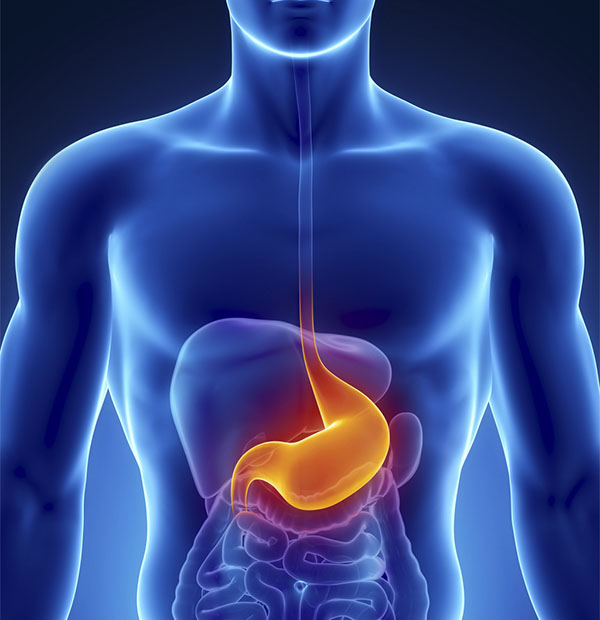
Capsule endoscopy is a procedure used to record internal images of the gastrointestinal tract for use in medical diagnosis. The capsule is similar in shape to a standard pharmaceutical capsule, although a little larger, and contains a tiny camera and an array of LEDs powered by a battery. After a patient swallows the capsule, it passes along the gastrointestinal tract taking a number of images per second which are transmitted wirelessly to an array of receivers connected to a portable recording device carried by the patient. The primary use of capsule endoscopy is to examine areas of the small intestine that cannot be seen by other types of endoscopy such as colonoscopy or esophagogastroduodenoscopy (EGD).

Dr Arti Rattan uses Capsule Endoscopy to find the cause of Gastrointestinal Bleeding which can lead to Iron Deficiency Anaemia, to diagnose IBD, to diagnose Celiac Disease and scan for polyps. Capsule endoscopy can also show tumors in the small intestine or other parts of the digestive tract.

Endoscopic ultrasound

Endoscopic ultrasound or echo-endoscopy is a minimally invasive medical procedure in which endoscopy is combined with ultrasound to obtain detailed images of the lining and walls of your digestive tract and chest, nearby organs such as the pancreas and liver, and lymph nodes. Dr Noureddin will use this procedure to evaluate cancer of the colon, oesophagus, pancreas or stomach, and Ampullary cancer, Lymphoma, Barrett’s Oesophagus, Neuroendocrine tumors, Pancreatitis and pancreatic cysts and Bile Duct Stones

Endoscopic Mucosal Resection

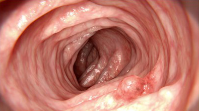
Gastrointestinal endoscopic mucosal resection (EMR) is a procedure used to remove abnormal tissue, tumors and other precancerous growths from the lining of the digestive tract without surgery.

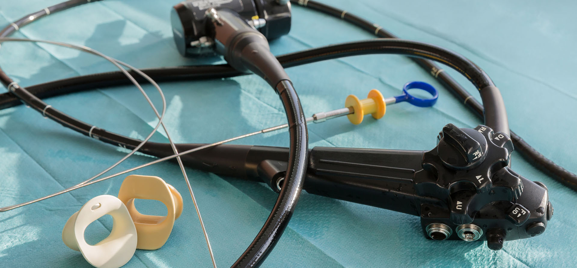
Endoscopic mucosal resection is performed using an endoscope, a long, narrow tube equipped with a light and video camera. During EMR of the upper digestive tract, the doctor passes the endoscope down your throat into your esophagus, stomach or upper part of the small intestine. For the colon and rectum, the doctor guides the tube up through the anus. Instruments are guided through this tube to remove growths or polyps. Because there is no incision, patients generally recover faster, with less pain than from regular surgery and can return home the same day.

EMR is usually done to treat a health condition. However, your doctor may also collect samples of tissue during the procedure. Examination of this tissue can help your doctor make a diagnosis. For example, if you have cancer, EMR can help determine if the cancer has spread to tissues beneath the digestive tract lining.


Disclaimer:

The information provided on this site is intended to educate the reader about certain medical conditions and certain possible treatment. It is not a substitute for examination, diagnosis, and medical care provided by a licensed and qualified health care professional. If you believe you, or someone you know suffers from the conditions described herein, please see your health care provider immediately. Do not attempt to treat yourself or anyone else without proper medical supervision.